

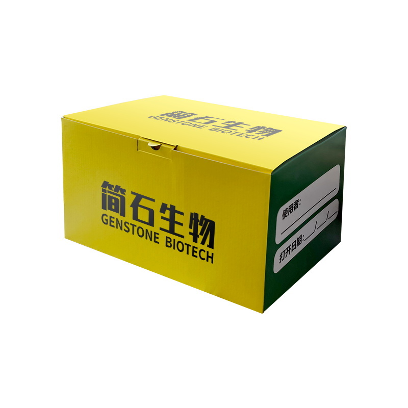
100 bp Standard DNA Ladder (TM1003)
| Cat # | Specifications | Unit | Price |
| TM1003-50 | 300 µL | support | ¥160 |
| TM1003-100 | 600 µL | support | ¥320 |
| TM1003-200 | 1200 µL | support | ¥550 |
Keywords
Product Description
FAQ
Product Information
Related Documents

TM1003 100 bp Standard DNA Molecular Weight Marker
DNA molecular weight standard with pre-mixed loading buffer, comprising seven bands ranging from 100 to 1500 bp, with a prominent 700-bp band, suitable for analyzing longer PCR products.
Core Features
- Wide coverage area : 100–1500 bp, suitable for longer fragments
- Highlight and locate : The 700-bp band becomes twice as bright, enabling rapid anchoring.
- Ready-to-use : Sample buffer preloaded
- Quantitative precision : The DNA quantity in the bands is fixed, allowing for semi-quantitative reference.
Strip information
- A total of 7 DNA fragments
- Fragment size: 100, 300, 500, 700, 900, 1200, 1500 bp
- 700 bp band : 100 ng per injection (highlighted)
- The remaining 6 bands : 50 ng per injection
Key Points of Use
- Recommended 1%–2% agarose gel
- Voltage: 4–10 V/cm
- Standard sample volume: 3–6 μl
- Compatible with conventional nucleic acid dyes
TM1003 Frequently Asked Questions
This product contains seven double-stranded DNA fragments, with sizes of 100 bp, 300 bp, 500 bp, 700 bp, 900 bp, 1200 bp, and 1500 bp, respectively. The 700-bp fragment is a bright band (100 ng per lane), while the remaining bands are each loaded at 50 ng per lane, facilitating rapid size determination and enhancing interpretation efficiency.
Recommended loading volume: 3–6 μl per lane;
Loading rule: For every 1 mm of well width, load 1 μl; if the wells are wider, slightly increase the sample volume to prevent overloading and subsequent band smearing.
This product comes pre‑mixed with loading buffer, so no additional preparation or addition is required. After thawing and mixing, it can be directly loaded onto the gel for electrophoresis, simplifying the experimental workflow.
Gel: 1–2% agarose gel, optimized for clear separation of DNA fragments ranging from 100 to 1500 bp;
Voltage: 4–10 V/cm (distance between the positive and negative electrodes). Excessively high voltage can cause gel overheating and band distortion, while excessively low voltage results in prolonged electrophoresis times.
The product concentration is approximately 67 ng/μl; based on a standard loading volume of 6 μl, the total DNA yield is about 402 ng, sufficient to meet the requirements for routine electrophoresis visualization and semi‑quantitative analysis.
Storage conditions: Store at low temperature; do not leave at room temperature for extended periods.
Note: Repeated freeze–thaw cycles can cause DNA fragmentation, blurred or missing bands, and uneven band intensity, directly compromising the accuracy of electrophoresis results. It is recommended to aliquot and store the sample as needed.
Buffer: Replace the electrophoresis buffer with a fresh one; ion imbalance in the old buffer can degrade separation performance.
Gel: Use freshly prepared agarose gels to avoid improper gel concentrations and uneven gelation.
Product: Avoid nuclease contamination and DNA degradation caused by repeated freeze–thaw cycles.
Operation: Control the sample loading volume and verify that the positive and negative terminals are not reversed.
This product is covered by a one-year quality warranty from the date of purchase, and all reagents have undergone rigorous quality testing. In the event of quality issues such as missing bands or degradation, please contact our after-sales service department with your proof of purchase for assistance.
Clip Range: TM1003 covers 100–1500 bp, while TM1002 covers only 100–700 bp.
Highlighted strip: TM1003 bright band at 700 bp, TM1002 bright band at 400 bp;
Applicable scenarios: TM1003 is suitable for identifying PCR products with longer fragments, while TM1002 is designed for rapid analysis of shorter fragments.
It must be used in conjunction with conventional nucleic acid dyes; the product itself does not contain any staining components.
After staining, visualize the sample using a UV gel imaging system to avoid DNA damage caused by prolonged UV exposure.
Tip: Use a 700-bp bright band as the exposure reference to prevent over- or under-exposure from affecting size determination.
document.addEventListener('DOMContentLoaded', function() { const faqItems = document.querySelectorAll('.faq-item'); faqItems.forEach(item => { const question = item.querySelector('.faq-question'); question.addEventListener('click', () => { // 只切换当前问题的展开状态,不关闭其他问题 item.classList.toggle('active'); }); }); // 移除了默认展开第一个问题的代码 // 移除了平滑滚动效果的代码 });
Related Products


Order Consultation: Hu Manager

Order Consultation: An Manager

End-user Scientific Research Customer Order Consultation

Technical Consultation: Bai Technical

Customer Service

Douyin

WeChat Video Account

Kuaishou

Contact Us
Jianshi Biotechnology (Beijing) Co., Ltd.
Company Address: Room 401, Building 11, No. 12 Courtyard, Juyuan Middle Road, Mapo Town, Shunyi District, Beijing
Phone: 86 18640171737
E-mail: gwang@jianshibio.com
Jianshi Biotechnology (Zhejiang) Co., Ltd.
Address: Building 11, No. 36 Xingshun Road, Zhongyi Ningbo Eco-park, Yuyao City, Zhejiang Province
WhatsApp: 86 18640171737
Leave a Message
If you are interested in our products, please leave your phone number.
Consult Now →Product Subscription
Subscribe to us to stay informed about product updates and special offers